
Figure 1.
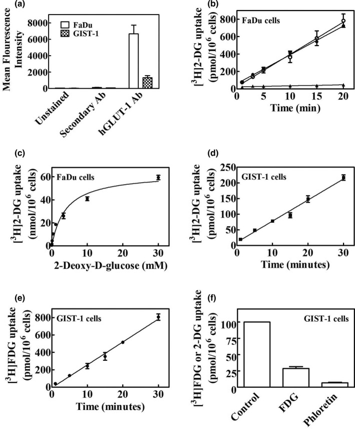
Analysis of hGLUT‐1 protein abundance and uptake of [3H]2‐DG and [3H]FDG in FaDU and GIST‐1 cells. (a) Flow cytometry analysis of hGLUT‐1 protein. FaDu and GIST‐1 cells were stained with hGLUT‐1 antibody for 60 minutes at RT followed by staining with secondary antibody at a dilution of 1/300. Cells were incubated for 30 minutes at RT in the dark. After washing, cells were analyzed for fluorescence by analytical flow cytometry. Mean results from three independent experiments with untreated cells and cells stained with secondary antibody only or hGLUT‐1 plus secondary antibody are shown. (b) [3H]2‐DG uptake in FaDu cells. Time courses of uptake of [3H]2‐DG in FaDu cells in presence of potassium or sodium with or without phloretin are shown (○, NaCl; ●, KCl; ▲, 200 µM Phloretin). (c) The concentration dependence (0–30 mM) of [3H]2‐DG uptake rates in FaDu cells. (d, e) Time courses of [3H]2‐DG and [3H]FDG uptake, respectively, in GIST‐1 cells. (f) Inhibition of [3H]2‐DG uptake by phloretin or excess FDG in GIST‐1 cells. Values with mean ± SE. are shown in each panel. All experiments were repeated three times with three to four replicates per condition. 2‐DG, 2‐deoxy‐d‐glucose; hGLUT, human glucose transporter; RT, room temperature.