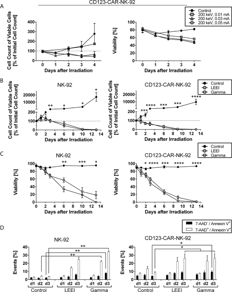
Figure 1.

Cell proliferation of NK-92 and CD123-directed CAR-NK-92 is fully inhibited by gamma irradiation and LEEI. (A) Cell count of viable CD123-CAR-NK-92 cells (left) and viability (right) were determined for 4 days for non-irradiated cells (black circles, n = 3) and LEE-irradiated cells at 200 keV and 0.01 mA (grey squares, n = 1), 0.03 mA (grey triangles, n = 3) or 0.05 mA (grey circles, n = 3) on an automated cell counter (CASY). (B) Cell count of viable NK-92 (left, n = 5) and CD123-CAR-NK-92 cells (right, n = 9) was determined for 13 days after irradiation for non-irradiated cells (black), LEE-irradiated cells (grey) and gamma-irradiated cells (white) on an automated cell counter (Countess II). (C) Viability was measured by trypan blue staining on an automated cell counter (Countess II) for 13 days after irradiation of NK-92 (left, n = 5) and CD123-CAR-NK-92 cells (right, n = 9). Non-irradiated cells (black) were compared to LEE-irradiated cells (grey) and gamma-irradiated cells (white). (D) Flow cytometry analysis of LEE- or gamma-irradiated NK-92 (left, n = 4) and CD123-directed CAR-NK-92 (right, n = 7) cells after 7-AAD and Annexin V staining. 7-AAD-/Annexin V+ (black) and 7-AAD+/Annexin V+ (white) subpopulations are shown. All values are indicated as means ± SEM, statistical significance is symbolized by asterisks (* for p ≤ 0.05, ** for p ≤ 0.01, *** for p ≤ 0.001, and **** for p ≤ 0.0001, ANOVA or Kruskal-Wallis test adjusted for multiple comparisons by Dunn’s test).
