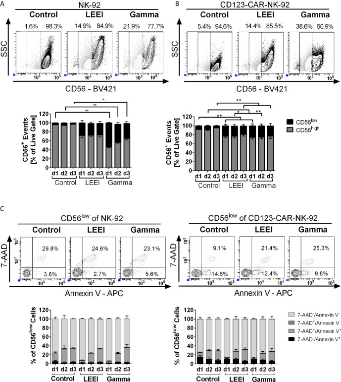
Figure 3.
Irradiation decreases surface expression of CD56. (A+B): NK-92 (A, n = 4) and CD123-CAR-NK-92 cells (B, n = 7) were stained with anti-human CD56 antibody. Surface expression levels of CD56 are indicated as CD56low (black) and CD56high (grey). Representative contour plots of non-irradiated (control), LEE-irradiated, and gamma-irradiated cells on day 3 post-irradiation are shown. (C) Analysis of the CD56low subpopulation of NK-92 (left) and CD123-CAR-NK-92 cells (right) regarding apoptosis: 7-AAD-/Annexin V- (lightest grey), 7-AAD+/Annexin V- (dark grey), 7-AAD+/Annexin V+(light grey) and 7-AAD−/Annexin V+ (black). Representative contour plots of non-irradiated (control), LEE-irradiated, and gamma-irradiated cells on day 3 post-irradiation are shown. Values are indicated as means ± SEM, statistical significance is symbolized by asterisks (* for p ≤ 0.05 and ** for p ≤ 0.01, Kruskal-Wallis test adjusted for multiple comparisons by Dunn’s test).

