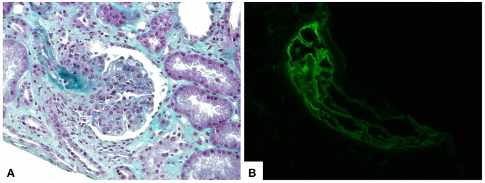
Figure 2.
Kidney biopsy showing lupus nephritis and thrombotic microangiopathy findings. (A) Histological sections demonstrate proliferative glomerulonephritis and fibrin thrombi in afferent arteriole (trichrome stain, 400×). (B) Immunofluorescence staining revealed fibrin deposition within the intravascular thrombi (fibrinogen antiserum, 400×).

