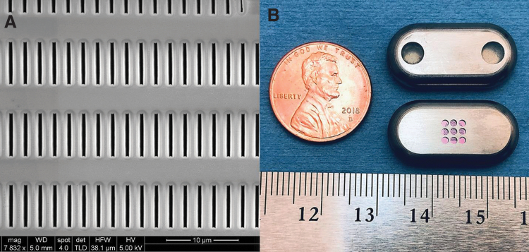
FIG. 3.
(A) Scanning electron microscopy image of the surface of the nanofluidic membrane, showing the inlet of the slit-nanochannels and the dense channel array. (B) Nanofluidic implant displaying the two loading and venting ports with self-sealing septa (top) and the nanochannel membrane (pink) assembled within the titanium drug reservoir (bottom).

