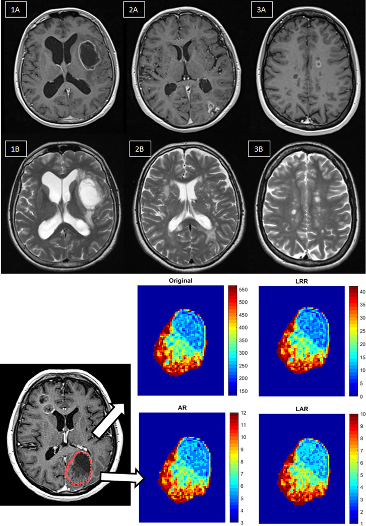
Fig 1.
a. Representative 3D T1 post-contrast (upper row) and T2-weighted axial (lower row) MR images for the selected disease groups. 1A-1B: Glioblastoma brain tumors, 2A-2B: Ischemia, 3A-3B: MS. b. Visualization steps of discretization workflow in radiomics. First, images are acquired. The next step is the segmentation of the region of interest, from which texture features are extracted. Then the discretization pre-processing steps are performed on the images, namely LRR, AR and LAR. The texture characteristics are then subtracted from the area of interest. The AR and LAR discretizations smooth the contrast much better within the segment, which is most easily observed at lower values (blue color).

