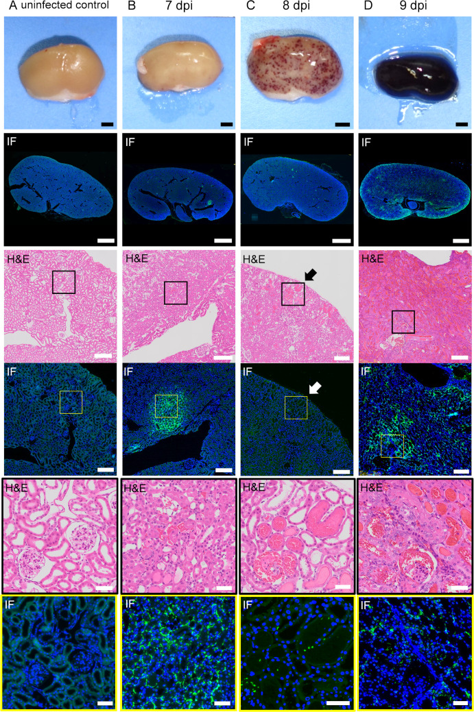
Fig 4. Distribution of leptospires and histopathological findings of infected kidneys in the acute phase.
The hamsters were subcutaneously injected with PBS (A) or 1 × 10⁴ cells of L. interrogans serovar Manilae strain UP-MMC-SM (B, C, D). The left kidneys collected from the moribund hamsters were fixed and sectioned into 4 μm slices, and then stained with immunofluorescent and H&E staining. Comparison of immunofluorescent staining and H&E staining of serial renal sections in the acute phase of infected hamsters. Representative macroscopic images and microscopic images of the serial sections are shown. Leptospires are labeled in green and cell nuclei in blue. The strong green fluorescence at the margin of the kidney is autofluorescence. (A): uninfected control, (B): 7 dpi, (C): 8 dpi, (D): 9 dpi. (B, C, D) Leptospires that colonize the kidneys of hamsters during the acute phase migrate through the interstitium and spread. (C) The section of petechiae on the kidney at 8 dpi was observed by H&E staining. The petechiae on the kidney (arrow) was a part of the hemorrhagic nephron-tubules. The areas boxed by black (third row) or yellow (fourth row) are enlarged in the fifth and sixth rows, respectively. The scale bars are 2 mm in the first and second rows; 200 μm in the third and fourth rows; and 100 μm in the fifth and sixth rows.

