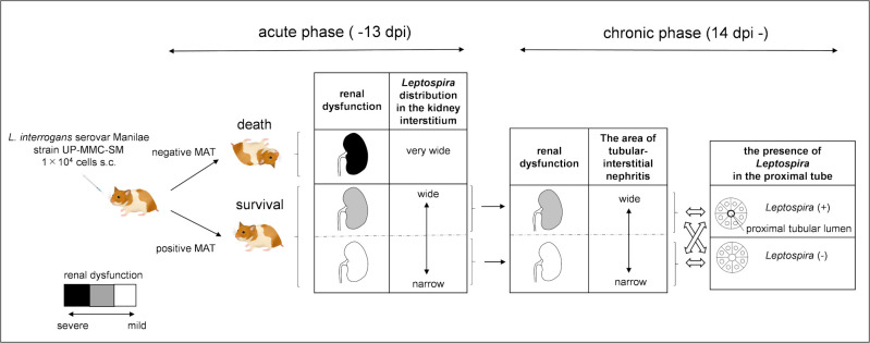
Fig 8. Hamster model of acute and chronic leptospirosis with subcutaneous infection.
The color scale of the longitudinal section of the kidney indicates the degree of renal dysfunction. Subcutaneous infection with pathogenic leptospires could cause acute death in hamsters, whereas recovery from acute infection led to chronic leptospirosis. If antibody production is initiated during the acute phase when leptospires colonize the kidney and proliferated there, the growth of leptospires is suppressed and hamsters survive. On the other hand, if antibody production is delayed, leptospires become overloaded and hamsters die. Leptospira distribution in the kidney of surviving hamsters was expected to be narrower than that of dead hamsters in the acute phase. The distribution of leptospires in the kidney during the acute phase may affect the chronic renal damage due to tubulointerstitial nephritis. Some hamsters showed leptospiral colonization in the renal proximal tubule lumen during the chronic phase, which was not correlated with elevated serum creatinine levels. The arrows (black) show the passage of time. The double-headed arrow (white) shows the relationship between renal dysfunction and the presence of Leptospira in the proximal tube.

