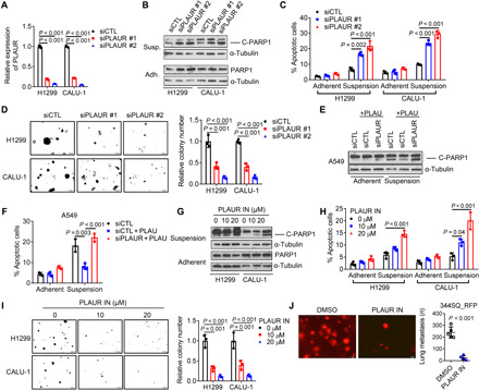
Fig. 5. Secreted PLAU inhibits anoikis through autocrine PLAUR activation.

(A) qPCR confirmation of PLAUR depletion in siRNA-transfected H1299 and CALU-1 cells. (B and C) Western blot analysis of cleaved PARP1 (B) and annexin V/propidium iodide flow cytometry (C) to detect apoptotic cells under adherent and suspension conditions. (D) Colonies formed in soft agar by siRNA-transfected cells. (E and F) Western blot analysis of cleaved PARP1 (E) and annexin V/propidium iodide flow cytometry (F) of siRNA-transfected cells treated with (+) or without recombinant PLAU (10 ng/ml) under adherent or suspension conditions. (G and H) Western blot analysis of cleaved PARP1 (G) and annexin V/propidium iodide flow cytometry (H) following treatment for 2 days with indicated concentrations of PLAUR inhibitor (PLAUR IN). (I) Colonies formed in soft agar following 14 days of PLAUR inhibitor treatment. (J) Lung metastases generated in syngeneic, immunocompetent mice by tail vein–injected RFP-tagged 344SQ cells that had been pretreated for 2 days with 10 μM PLAUR inhibitor or vehicle [dimethyl sulfoxide (DMSO)]. Results represent means ± SD values from a single experiment incorporating biological replicate samples (n = 3, unless otherwise indicated) and are representative of at least two independent experiments carried out on separate days. P values, two-tailed Student’s t test for two-group comparisons and one-way ANOVA test for multiple comparisons.
