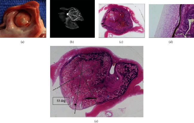
Figure 3.

CLAAS gross and histological findings: (a) photograph of the gross appearance of the CLAAS device from the left atrium showing complete healing of the implant which was covered by a thin neointima and complete seal of the LAA ostium. (b) A/P view of endoskeleton. (c, d) Sagittal hematoxylin and eosin- (H&E-) stained sections showing complete occlusion of the LAA ostium. Also noted is the absence of thrombus or signs of inflammation. (e) H&E stain showing complete seal even with off-axis positioning of the implant. The LAA is filled with a mixture of the device material, fibrous connective tissue, and minimal residual thrombus. The ostium of the LAA is covered by thin neointima comprised of fibrous connective tissue.
