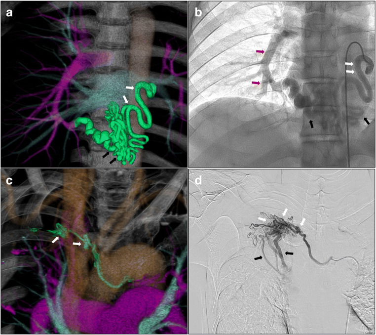
Fig. 1.
Three-dimensional CT (a, c) and angiography (b, d) findings before BAE. a Abnormal pulmonary ligament artery with vessel dilation (white arrows) and tortuosity (black arrows). b Vessel dilation (white arrows), tortuosity (black arrows), and systemic artery-pulmonary artery direct shunting (red arrows). c Abnormal bronchial artery with hypervascularity (white arrows). d Hypervascularity (white arrows) and systemic artery-pulmonary artery direct shunting (black arrows)

