Fig. 2.
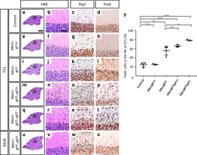
Mert::Bfl/fl::Nfl/wt and Mert::Bfl/fl::Nfl/fl mice display increased numbers of Pax6 positive cells in the molecular layer. Representative sagittal H&E sections of whole cerebella are shown at P21 and at adult age (a, e, i, m, q and u). Brg1 knockout (c, g, k, o, s, w) is examined by IHC. Sagittal H&E (b, f, j, n, r, v) and Pax6 (d, h, l, p, t, x) stainings of the EGL and IGL display an aggregation of Brg1 deficient granule cells. Quantification of Pax6 expressing cells in the ML of animals at P21 are shown in Y. The control group includes Bfl/fl::Nfl/wt and Bfl/fl::Nfl/fl mice (n = 4). The mutant groups (Mert::Bfl/wt, Mert::Bfl/fl, Mert::Bfl/fl::Nfl/wt and Mert::Bfl/fl::Nfl/fl) include 3 animals each. The scale bar in A corresponds to 400 μm and is representative for e, i, m, q, and u. The scale bar in B corresponds to 50 μm and is representative for all other panels. **p < 0.01, ***p < 0.001, ****p < 0.0001. n.s., not significant

