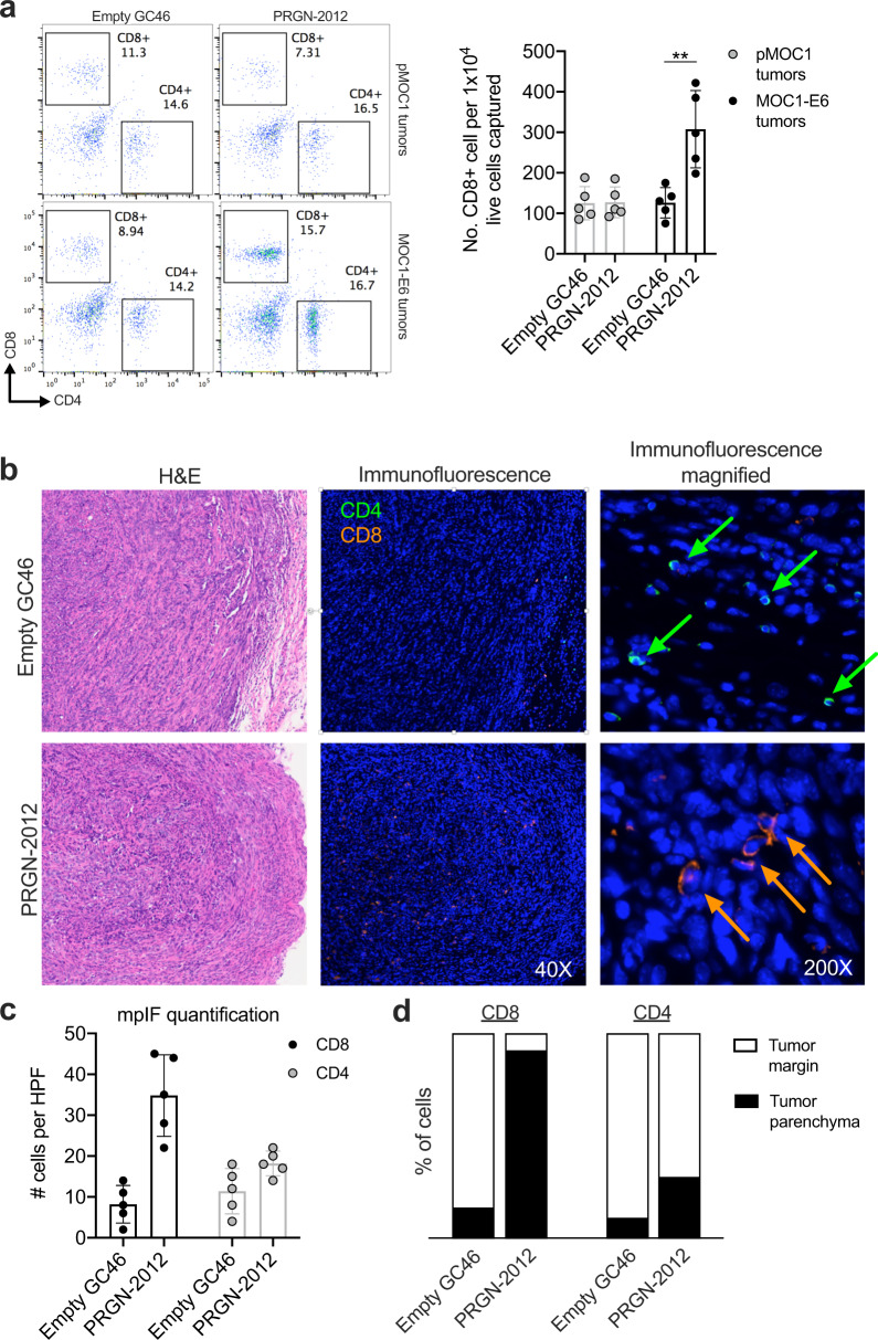
Fig. 5. PRGN-2012 promoted trafficking of CD8+ T lymphocytes into the tumor parenchyma.
a Representative dot plots of freshly digested parental pMOC1 or MOC1-E6 tumors obtained from mice treated with PRGN-2012 or empty GC46 assessed for T-lymphocyte accumulation via flow cytometry. Normalized quantification is shown on the right. b Representative H&E and immunofluorescence photomicrographs demonstrating localization of T lymphocytes within MOC1-E6 tumors treated with PRGN-2012 or empty GC46. c Quantification of total T lymphocytes per high power field (HPF). mpIF multiplex immunofluorescence. d Quantification of tumor margin vs tumor parenchyma localization of CD8+ and CD4+ T lymphocytes.

