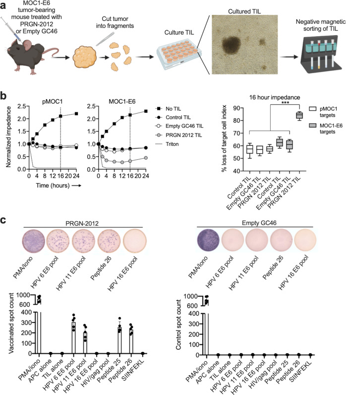
Fig. 6. HPV antigen-specific effector T lymphocytes infiltrated tumors following PRGN-2012 treatment.
a Schematic of TIL cultured from tumors from MOC1-E6 tumor-bearing mice treated with PRGN-2012 or empty GC46 vector (n = 5 mice/group). b Representative impedance analysis of cultured TIL from MOC1-E6 tumors treated with PRGN-2012, empty GC46, or control (PBS), then cocultured with either parental MOC1 or MOC1-E6 target tumor cells (n = 5 tumors per condition). TIL are added to target cells at experimental time 0. Percent killing (represented as % loss of target cell index) is quantified 16 h after the addition of TIL to targets and is shown on the right. c Photomicrographs of representative ELISpot wells and quantification of IFNγ spots demonstrating responses to HPV6 and 11 overlapping 15-mer peptide pools as well as synthesized minimal peptides in TIL cultures from MOC1-E6 tumors treated with PRGN-2012 or empty GC46. TIL tumor-infiltrating lymphocytes. ***p < 0.001; ANOVA.

