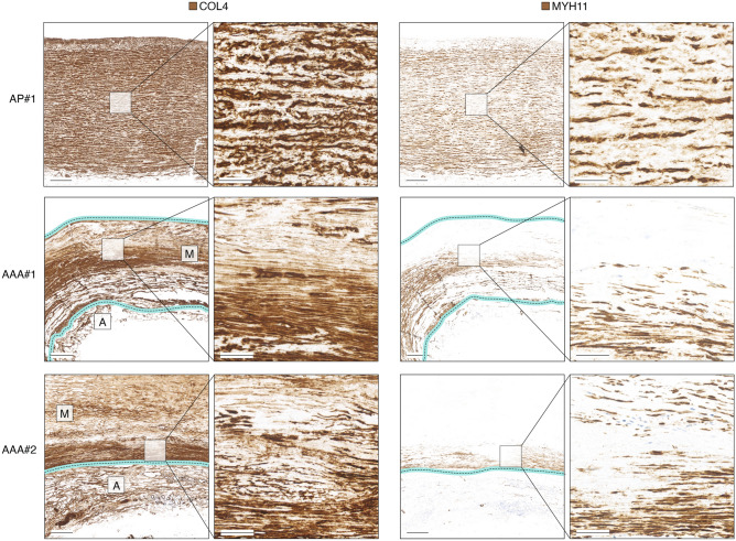
Figure 4.
COL4 and MYH11 immunohistochemical staining of non-lesioned ascending aorta and two examples of AAA lesions. To assist assessment of AAA lesions, the partially degraded medial (M) and adventitial (A) layers are labeled on overview images of COL4 stainings, and the media borders are marked with cyan/dotted lines. Images are representative specimens of 5 aortic punch- and 5 AAA-samples analyzed, and images of remaining specimens are shown in Supplementary Fig. 5. Scalebars (overview) = 1 mm; Scalebars (magnification) = 200 µm. AP = Aortic punch (ascending thoracic aorta). #1–#2 refer to sample IDs.

