Figure 3.
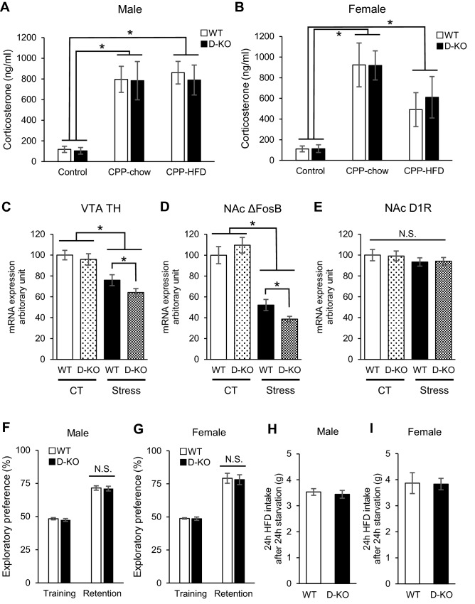
Caloric restriction affected the mRNA expressions of TH in VTA and ΔFosB in NAc via GR signaling in dopaminergic neurons, while corticosterone levels, cognitive function, and refeeding induced by caloric restriction stress showed no differences. (A,B) Serum corticosterone levels at the end of the CPP session, compared to control (under no stress). Both D-KO and WT exhibited significantly higher levels of serum corticosterone, with no difference between genotypes nor between feeding conditions (A: male, B: female, n = 5–7 for each group). (C–E) mRNA expressions of TH in VTA (C), ΔFosB (D) and D1R (E) in NAc after caloric restriction (termed ‘Stress’). In the control group, there were no significant differences in the mRNA expression levels between genotypes (C–E). The caloric restriction reduced the mRNA expressions of TH and ΔFosB in both D-KO and WT mice compared to control groups, with significant reduction in D-KO compared to WT (C,D) (female mice, n = 7–10 for each group). (F,G) NORT results. There were no significant differences in exploratory preference between D-KO and WT in both the training and retention sessions (F: male, G: female, n = 9–13 for each group). (H,I) HFD refeeding after fasting for 24 h. Both D-KO and WT, male (H) and female (I) mice exhibited no significant differences in the amount of HFD consumed (n = 5–7 for each group). All values are mean ± SEM. Statistical analyses were performed using two-way factorial ANOVA followed by Bonferroni post hoc test (A-G) or unpaired t test (H, I). Asterisks denote significant group differences at p < 0.05 vs control for (A-D).

