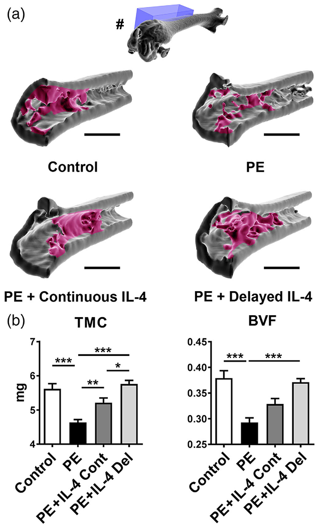
FIGURE 1.

The effects of IL-4 delivery on the particle-induced osteolysis as assessed by μCT. Polyethylene (PE) particles were delivered into the mouse distal femur for 8 weeks through hollow titanium rod. IL-4 was added to the particle infusion either from the beginning of the particle delivery (IL-4 continuous, IL-4 Cont) or after 4 weeks (IL-4 delayed, IL-4 Del). At 8 weeks, the amount of bone at the distal femur was determined by μCT imaging. (a) 3D reconstructions of a sagittal segment of distal metaphyseal region of the femur. Note the loss of metaphyseal and peri-implant trabecular bone in the PE infused samples and the restoration of bone in the IL-4 treated samples. In the 3D reconstruction of the whole femur (#) the location of the sagittal 3D reconstructions (blue box) is shown. Notice also the rod channel visible in the intercondylar region of the whole femur reconstruction. (b) Bar diagrams showing the tissue mineral content (TMC) and bone volume fraction (BVF) at the distal femur. n = 12 mice per group. * = p < .05, ** = p < .01 *** = p < .001. Main regions of metaphyseal trabecular bone are indicated with red. Scale bars 1,500 μm
