
FIGURE 2.
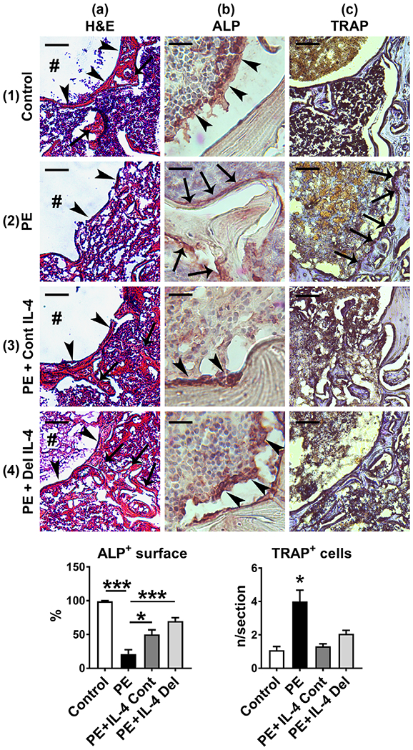
The effects of IL-4 delivery on the bone microstructure and the amount of osteoclasts and osteoblasts. Polyethylene (PE) particles were delivered into the mouse distal femur for 8 weeks through hollow titanium rod. IL-4 was added to the particle infusion either from the beginning of the particle delivery (IL-4 continuous, IL-4 Cont) or after 4 weeks (IL-4 delayed, IL-4 Del). At 8 weeks, the overall tissue morphology at the distal femur was evaluated from hematoxylin and eosins (H&E) stained transverse tissue sections (panels 1–4a). In the control sample note the formation of a bony interface (arrowheads) around the end of the implant channel (#), surrounded by a network of trabecular bone (arrows) and bone marrow (panel 1a). Note also the loss of bony interface (arrowheads) and the surrounding trabecular bone in the PE treated sample (panel 2a) and the marked restoration of these bone structures in the IL-4 infused samples (panels 3–4a, arrowheads and arrows). The amount of osteoblasts was determined by ALP immunostaining (Panels 1-4b the left bar diagram, * = p < .05, *** = p < .001). Note the large cuboidal osteoblasts (brown staining cells, arrowheads) covering the endosteal surfaces of the bone trabecula in the control and in the IL-4 treated samples. In contrast, in the PE sample endosteum was either devoid of ALP positive cells or covered by flat inactive appearing bone lining cells (arrows). The amount of osteoclasts was determined by TRAP histochemistry (panels 1–4c and right bar diagram, * = p < 0.05 PE vs. all other groups). Note the increase in the amount of osteoclasts and the marked erosion of the bone in the PE sample (purple staining cells, arrows) and the reduction of the osteoclast number by both of the IL-4 treatments. n = 4 mice per group. Scale bars in the panels 1–4a 100 μm; Panels 1–4b 20 μm; Panels 1–4c 50 μm. ALP, alkaline phosphatase; TRAP, tartrate resistant acid phosphatase