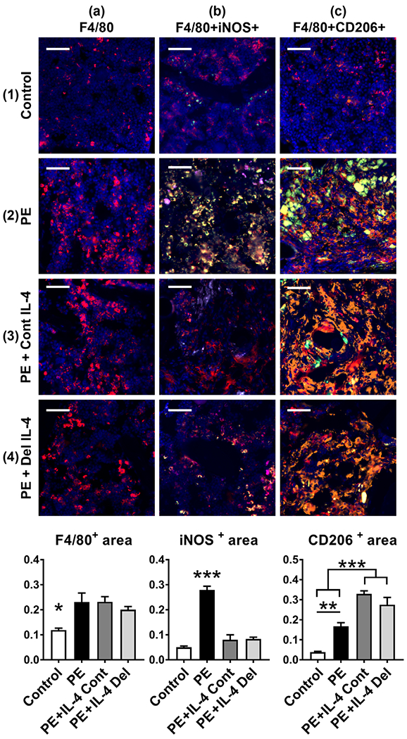
FIGURE 3.

The effects of IL-4 delivery on the macrophage polarization. Polyethylene (PE) particles were delivered into the mouse distal femur for 8 weeks through hollow titanium rod. IL-4 was added to the particle infusion either from the beginning of the particle delivery (IL-4 continuous, IL-4 Cont) or after 4 weeks (IL-4 delayed, IL-4 Del). At 8 weeks, the amount of macrophages at the distal femur was determined by F4/80 immunofluorescence staining (panels 1–4a, left bar diagram, * = p < .05 control vs. all other groups). Notice the increase in the amount of F4/80 positive macrophages in the metaphyseal bone marrow with the PE treatment. The amount of M1 macrophages was determined by F4/80 iNOS immunofluorescence double staining (panels 1–4b, middle bar diagram, *** = p < .001 PE vs. all other groups). Notice the increase of F4/80 + iNOS double positive M1 macrophages in the PE sample and the reduction in the amount of M1 macrophages by both of the IL-4 treatments. The amount of M2 macrophages was determined by F4/80 CD206 immunofluorescence double staining (panels 1–4c, right bar diagram, ** = p < .01 *** = p < .001). Notice the increase of F4/80 + CD206 double positive M2 macrophages by both of the IL-4 treatments. A modest increase in the M2 macrophages was observed also in the PE samples. F4/80—red; iNOS—green; CD206—orange; DAPI—blue. The large green bodies visible in some of the pictures are polyethylene particle aggregates. n = 4 mice per group. Scale bars 50 μm
