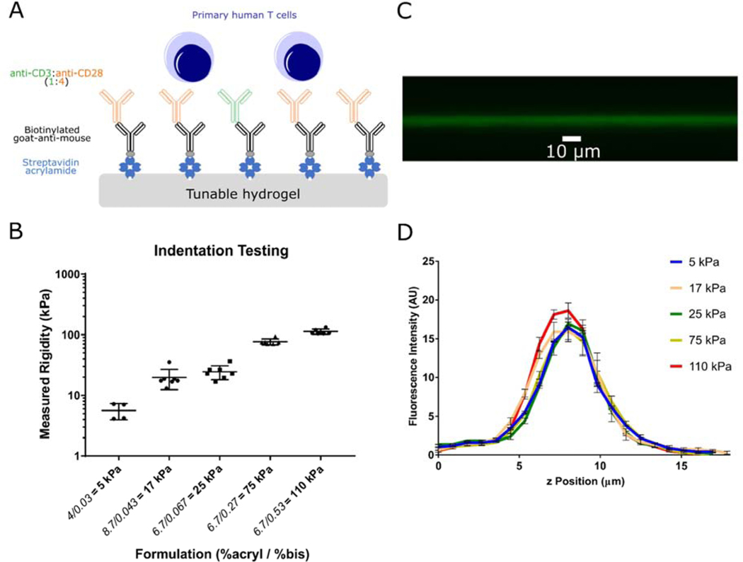
Figure 1. Characterization of the hydrogel system for T cell activation.
(A) Schematic of antibody coated hydrogels as T cell activation platform. Polyacrylamide gels were polymerized with streptavidin-acrylamide, then coated with biotinylated Goat-anti-mouse and secondary activating antibodies to CD3 and CD28. Mixed CD4+/CD8+ polyclonal primary human T cells were stimulated on activating substrates. (B) Mechanical testing via indentation indicate hydrogels with varying ratios of acrylamide and bis-acrylamide have Young’s modulus between 5 to 110 kPa. (Data are mean ± s.d., n=4 gels for 5 kPa, n=7 gels for all other formulations) (C) Confocal microscopy imaging of fluorescent antibodies coated on hydrogels indicate attachment of antibodies to hydrogel surface. (D) Streptavidin-acrylamide concentrations were varied to obtain similar coating of antibodies on hydrogels, as verified by fluorescence intensities on the surface of gels. (n=3 gels for each formulation)

