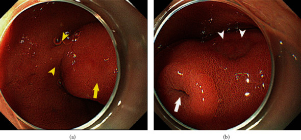
Figure 1.

(a, b) Duodenoscopy showing a pedunculated (arrowheads in (a)), polypoid, 4 cm mass at the duodenal bulb. A secretory portion at the top of the tumor is visible (arrow in (b)) as well as an ulcerated lesion without bleeding (arrow in (a) and arrowheads in (b)).
