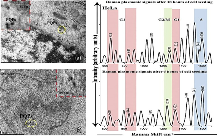
Fig. 24.
a The Bio-TEM image showing the presence of PQPs inside the cancerous cells (HeLa), b Bio TEM images show the interaction between plasmonic probes and nucleus components. c Plasmonic Raman signals response for cervical cancer (HeLa) after 6 and 18 h of cell seeding to monitor the growth of cells by studying the G0 G1 and G3 phase of the cell cycle [123]

