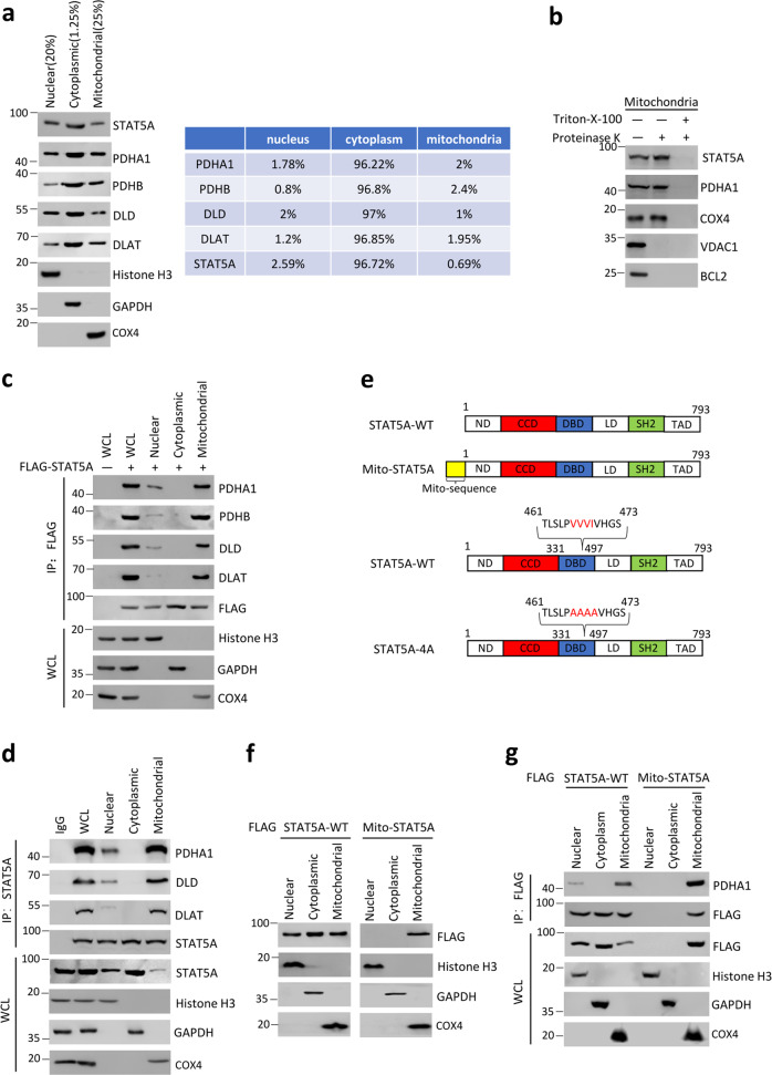
Fig. 2. The mitochondria is a main cellular compartment that STAT5A–PDC interaction occurs.
a The cytoplasmic, mitochondrial, and nuclear fractions from HeLa cells were prepared as described in the “Materials and methods” section. STAT5A and PDC subunit (PDHA1, PDHB, DLD, and DLAT) in the three fractions were detected by immunoblot. Histone H3 (nucleus), GAPDH (cytoplasm), and COX4 (mitochondria) were used as subcellular fraction markers. The total proteins used for subcellular fractionation were 4–5 mg. b Mitochondria were fractionated from HeLa cells and incubated with (lanes 2 and 3) or without (lane 1) proteinase K. To disrupt mitochondrial integrity, Triton X-100 was added in the digestion buffer (lane 3). The indicated proteins in the reactions were detected by Immunoblot. The total proteins used for isolation of mitochondria were 4–5 mg. c The nuclear, cytoplasmic, and mitochondrial fractions from HeLa cells transfected with expression vectors encoding EV or FLAG-STAT5A. WCL were immunoprecipitated with anti-FLAG antibody, and the immunoprecipitated proteins were immunoblotted with indicated antibodies. d The nuclear, cytoplasmic, and mitochondrial fractions from HeLa cells were subjected to immunoprecipitation with anti-STAT5A antibody, and the immunoprecipitated proteins were immunoblotted with indicated antibodies. e Schematic diagram of STAT5A-WT, Mito-STAT5A, and STAT5A-4A mutant. f The nuclear, cytoplasmic, and mitochondrial fractions from HeLa cells transfected with indicated expression vectors. The fractionated proteins were immunoblotted with indicated antibodies. g HeLa cells were transfected with expression vectors encoding FLAG-STAT5A-WT or FLAG-Mito-STAT5A.WCL were immunoprecipitated with anti-FLAG antibody, and the immunoprecipitated proteins were immunoblotted with indicated antibodies.

