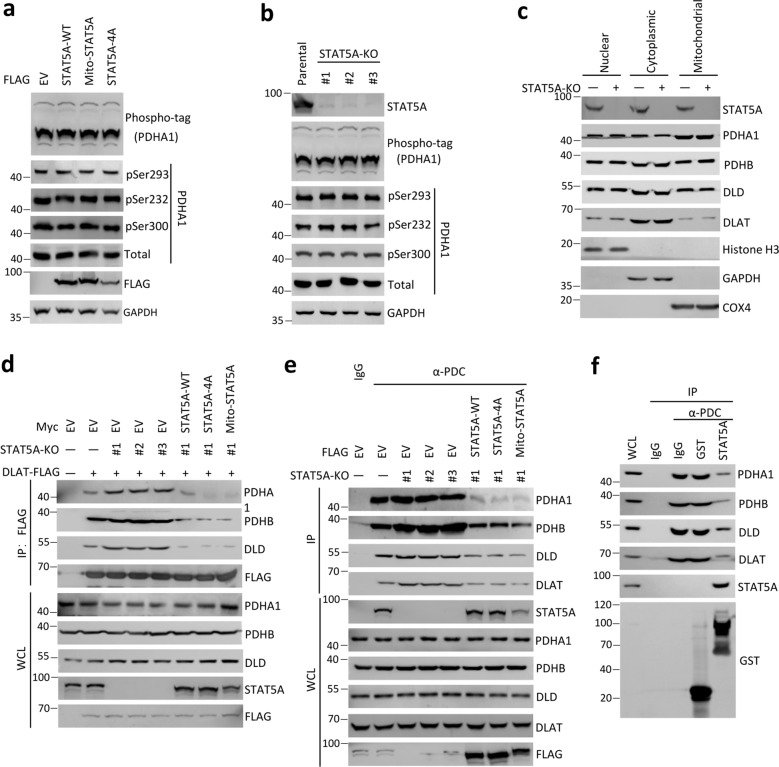
Fig. 4. Mitochondrial STAT5A suppresses PDC formation.
a HeLa cells were stably transfected with indicated expression vectors. The WCL were immunoblotted with indicated antibodies. The phosphorylated level of PDHA1 (p-PDHA1) was detected by phos-tag SDS-PAGE using an anti-PDHA1 antibody. b Immunoblots of the indicated proteins in WCL from parental or STAT5A-KO HeLa cells using CRISPR/Cas9 methods. The phosphorylated level of PDHA1 (p-PDHA1) was detected by The WCL were immunoblotted with indicated antibodies. Phos-tag SDS-PAGE was performed using an anti-PDHA1 antibody. Parental HeLa cells were used as the control. c The nuclear, cytoplasmic, and mitochondrial fractions from parental and STAT5A-KO HeLa cells. The fractionated proteins were immunoblotted with indicated antibodies. d STAT5A-KO HeLa cells were stably transfected with indicated expression vectors. Then, the cells were transiently transfected with expression vectors encoding DLAT-FLAG. WCL were immunoprecipitated with anti-FLAG antibody, and the immunoprecipitated proteins were immunoblotted with indicated antibodies. e STAT5A-KO HeLa cells were transfected with indicated expression vectors. Cell lysates were immunoprecipitated with anti-PDC immunocapture antibody, and the proteins were immunoblotted with indicated antibodies. IgG was used as a negative control for Immunoprecipitation. f The intact PDC was immunoprecipitated from HeLa cells by anti-PDC immunocapture antibody and then incubated with the indicated recombinant proteins (GST or GST-STAT5A, 2 μg) for 12 h. Bound materials and recombinant proteins were detected by Immunoblot.

