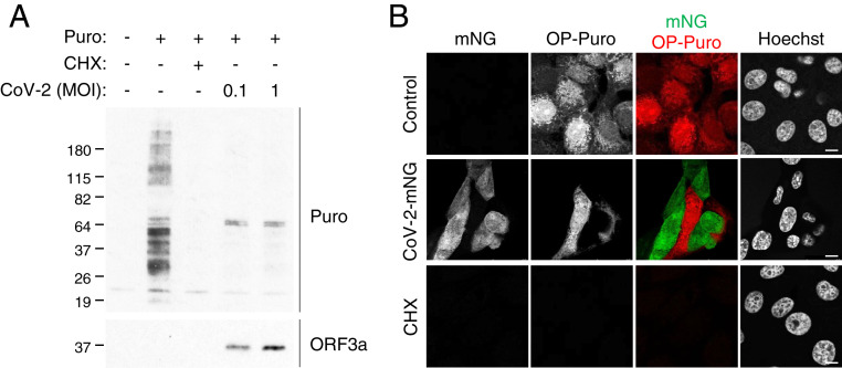
Fig. 1.
SARS-CoV-2 infection inhibits cellular translation. (A) Vero E6 cells were infected with SARS-CoV-2 at the indicated MOIs. After 24 h of infection, cells were pulse labeled with puromycin for 15 min. Puromycin incorporation was determined by immunoblotting using anti-puromycin antibody (Puro). Expression of SARS-CoV-2 viral protein was determined using anti-SARS-CoV ORF3a antibody (ORF3a). (B) Confocal images of Vero E6 cells infected with recombinant SARS-CoV-2 expressing mNeonGreen (CoV-2-mNG) (22) at an MOI of 0.5. After 24 h of infection, cells were pulse labeled with OP-Puro for 1 h, fixed, fluorescently labeled by the Click chemistry reaction, and stained by Hoechst. (Scale bars, 10 µm.)

