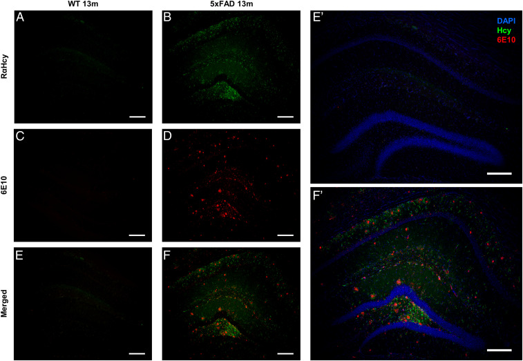
Fig. 5.
Detection of Hcy fibrils correlates with β-amyloid1-42 aggregation in AD model mice. The images represent sections of 5xFAD or WT animals. (A and B) Sections stained with anti–Hcy-fibrils antibodies (green). (C and D) Sections stained with anti–β-amyloid1-42 antibody 6E10 (red). (E and F) Merge of Hcy and β-amyloid1-42 staining. (E’ and F’) Enlarged images of E and F, respectively, merged with DAPI staining. (Scale bar, 200 µm.)

