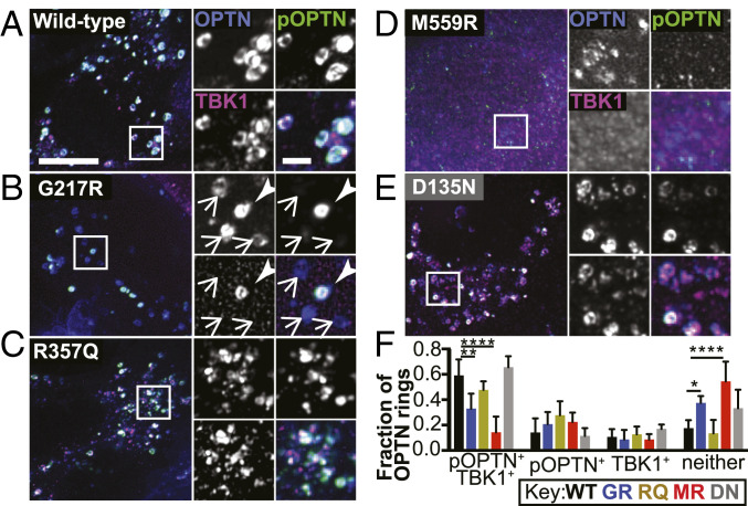
Fig. 5.
ALS-linked TBK1 mutants suppress OPTN phosphorylation. (A–E) Maximum intensity projection images of HeLa cells depleted of endogenous TBK1 and expressing Parkin (not tagged), OPTN (blue), and WT- (A), G217R- (B), R357Q- (C), M559R- (D), or D135N- (E) TBK1 variants (magenta), fixed after treatment with CCCP for 90 min. Phospho-S177 OPTN is tagged with an antibody (green). In B, one ring is positive for phospho-OPTN and TBK1 (arrowhead), and the others are negative for both (arrows). (Scale bars: zoom out, 10 μm; zoom in, 2 μm.) Images shown are insets; for representative images of whole fields, reference SI Appendix, Fig. S6A. (F) For each cell, the percentage of OPTN rings in each category was calculated. Error bars indicate SD. n = 8 to 15 cells from at least three independent experiments. *P ≤ 0.05, **P < 0.01, ****P < 0.0001 by ordinary one-way ANOVA with Dunnett’s multiple comparisons test.

