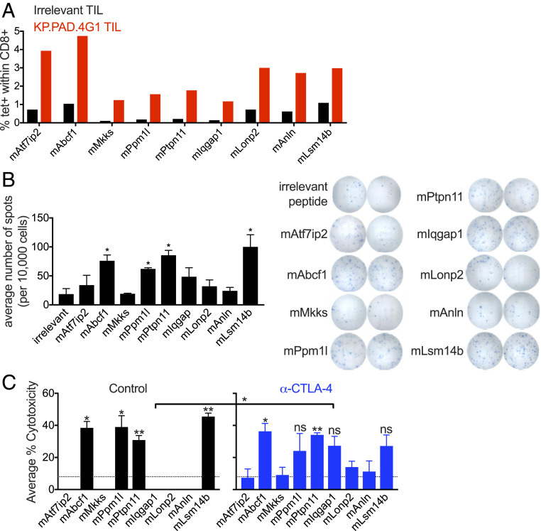
Fig. 3.
Tumor cell clone KP.PAD.4G1 contains unique radiation-induced mutations that lead to de novo antitumor CD8+ T cell responses. (A) TIL was isolated from KP.PAD.4G1 and irrelevant MCA-induced T3 tumors 11 d post transplantation and representative quantification of H-2-Kb p-MHC-I tetramer staining is shown as the percentage of CD8+ T cells of viable CD45+ cells within the tetramer-positive gate for the indicated KP.PAD.4G1 irradiation-induced mutations. (B) CD8+ T cells were enriched from KP.PAD.4G1 TIL and stimulated with splenocytes pulsed with the indicated peptides. IFNγ secretion was measured using ELISpot. Representative data are shown as the average number of spots per 10,000 CD8+ T cells ± SEM. (C) LDH cytotoxicity ELISA from splenocytes at day 12 post KP.PAD.4G1 tumor inoculation in control mAb-treated (black) and anti–CTLA-4–treated (blue) mice. Data represent the average percent cytotoxicity from two independent biological repeats ± SEM. Asterisks above bars indicate significance compared with the level of cytotoxicity observed against an irrelevant antigen (denoted by dashed lines). ns, not significant; *P < 0.05, **P < 0.01.

