Fig. 3.
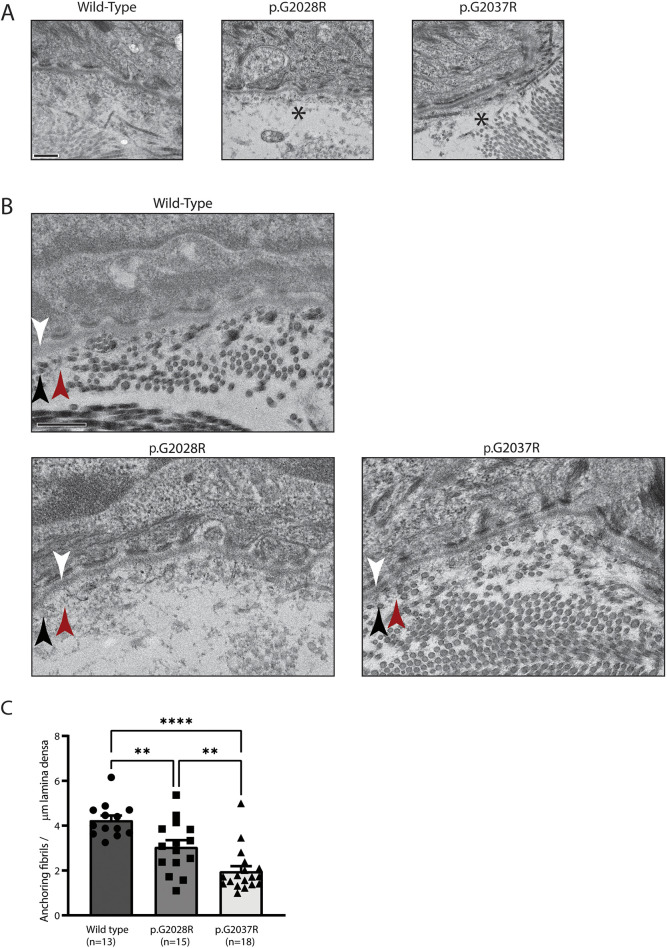
Electron microscopy reveals putative separation and reduced anchoring fibrils. (A) Electron microscopy of 12-week-old back skin shows putative separation below the lamina densa of the epidermal basement membrane, indicated by asterisks. Data are representative of multiple fields from a single biological replicate. Scale bar: 400 nm. (B) Electron microscopy also shows possible changes to the anchoring fibrils (indicated by red arrowheads) that sit below the lamina densa (indicated by white arrowheads) and among the interstitial collagen fibres (indicated by black arrowheads). Data are representative of multiple fields from a single biological replicate. Scale bar: 400 nm. (C) Quantification of anchoring fibrils per µm lamina densa, showing a reduction in both mutant mice. Number of random fields per genotype is indicated in the figure. Analysis by one-way ANOVA, **P<0.01, ****P<0.0001.

