Fig. 4.
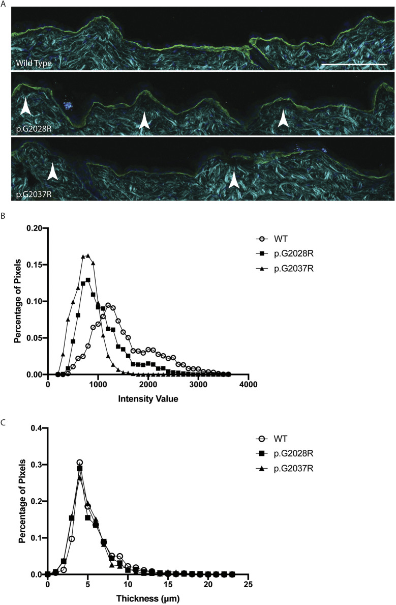
Immunofluorescence staining shows changes in both collagen VII expression and basement membrane structure. (A) Immunofluorescent staining of collagen VII (green) and DAPI (dark blue) in conjunction with second-harmonic imaging (cyan), which shows the structure of ordered collagens, in sections from 12-week-old mouse back skin. Changes in the intensity and thickness of collagen VII in the mutant sections are apparent when compared with wild-type skin, as is the presence of micro blisters (indicated by white arrowheads) below the basement membrane. Scale bar: 100 μm. (B) Analysis of the stained sections (five images per replicate, from three biological replicates per genotype) shows that the intensity of collagen VII staining varies across the mutants, with p.G2028R, and p.G2037R showing slightly less intensity than wild type (WT). (C) Analysis of the thickness of collagen VII staining shows that the p.G2028R and p.G2037R have a similar thickness to wild type.

