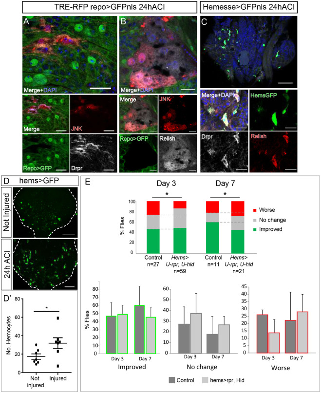
Fig. 7.
Phagocytes are recruited to injured VNCs and required for functional recovery. (A) Surface of metathoracic neuromere (MtN) stained for Drpr (grey) and active JNK signaling (TRE-RFP, red) in non glial cells. Glial cells (repo>GFP) are in green; nuclei were stained with DAPI (blue). The top image is a magnified version of the merged image below. (B) Surface of MtN showing non glial cells stained for Relish (grey) and active JNK signalling (red); nuclei were stained with DAPI (blue). The top image is a magnified version of the merged image below. (C) MtN hemocytes stained for Drpr (grey) and Relish (red); nuclei were stained with DAPI (blue). The boxed area is shown magnified in the four images at the bottom in A-C. (D) Representative images and quantification of hemocytes in not injured and injured MtNs. (D′) Plotted are the number of hemocytes in not injured and injured MtNs. Unpaired t-test, *P<0.05 (E) Quantification of the functional recovery. Percentage of flies whose hemocytes had been genetically ablated compared to that of control flies. Data were quantified for each category of movement, i.e. Worse (red), No change (grey) or Improved (green). Chi-square test with two degrees of freedom, *P<0.1. Genotypes: TRE-RFP; repoGal4>UAS-GFPnls (A,B). HemesseGal4>UAS-CD8GFP (C,D), CD8 is a transmembrane protein, therefore the cell surface is labeled with GFP. HemesseGal4>UAS-RedStinger (D′). UAS-reaper, UAS-hid (Control) and tubGal80ts; HemesseGal4>UAS-reaper, UAS-hid (E). Scale bars: 15 μm (A, B, C bottom image); 50 μm (C top image, D).

