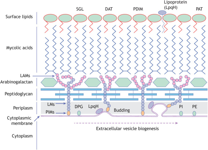
Fig. 3.
Mycobacterium tuberculosis cell wall. Diagram of the mycobacterial cell wall, indicating the location of key lipid components and the site of membrane budding to form EVs. DAT, diacyltrehalose; DPG, diphosphatidylglycerol; LAMs, lipoarabinomannans; LM, lipomannan; PAT, polyacyltrehalose; PDIM, phthiocerol dimycocerosate; PE, phosphatidylethanolamine; PI, phosphatidyl-myo-inositol; PIMs, phosphoinositol mannosides; SGL, sulfoglycolipid.

