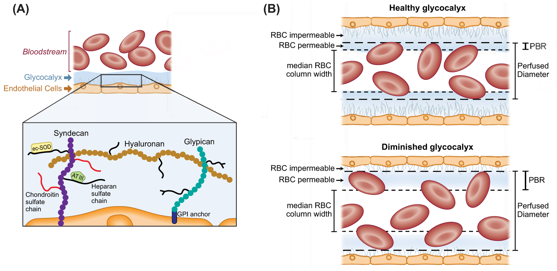
Figure 4. Schematic representation of the endothelial glycocalyx.

(A) Left side illustrating microvascular endothelial glycocalyx components including the network of core proteoglycans syndecan-1, with covalently bound long unbranched glycosaminoglycan side-chains (GAGs) heparin sulfate and chondroitin sulfate, noncovalently bound hyaluronan, and glypican with short-branched carbohydrate side chains. Also depicted are the dynamic layer of plasma proteins, including extracellular superoxide dismutase (ecSOD) and antithrombin III (AT III) that loosely bind with proteoglycans and glycosaminoglycans promoting protective functions of the endothelium and create a cross-linked mesh providing further stability to the glycocalyx layer. Syndecans bind the plasma membrane via a transmembrane domain and glypicans bind via a glycosylphosphatidylinositol (GPI) anchor. (B) Right side, illustrating a healthy endothelial glycocalyx which is relatively impermeable to red blood cells (RBCs) and other circulating cells in the microcirculation (right, upper panel), whereas a diminished glycocalyx allows for deeper penetration of RBCs into the glycocalyx layer (right, lower panel). This greater penetration of RBCs into the glycocalyx can be quantified in vivo as an increase in the perfused boundary region (PBR) in sublingual microvessels between 5 and 25 μm in diameter in humans. A larger PBR reflects a diminished or thinner glycocalyx layer compared with a smaller PBR and thicker glycocalyx.
