
FIGURE 1.
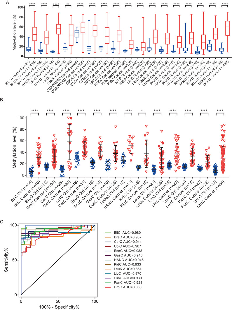
Hypermethylated PCDHGB7 is identified as a UCOM marker. (A) PCDHGB7 was hypermethylated in 17 cancer types compared with their normal tissues in TCGA databases. Box and whiskers plots were plotted; box represents the upper quartile, lower quartile, and median; whiskers represent minimum to maximum. BLCA, bladder urothelial carcinoma; BRCA, breast invasive carcinoma; CESC, cervical squamous cell carcinoma and endocervical adenocarcinoma; CHOL, cholangiocarcinoma; COADREAD, colon adenocarcinoma and rectal adenocarcinoma; ESCA, esophageal carcinoma; GBM, glioblastoma multiforme; HNSC, head and neck squamous cell carcinoma; KIRC, kidney renal clear cell carcinoma; KIRP, kidney renal papillary cell carcinoma; LIHC, liver hepatocellular carcinoma; LUAD‐LUSC, lung adenocarcinoma and lung squamous cell carcinoma; PAAD, pancreatic adenocarcinoma; PRAD, prostate adenocarcinoma; SARC, sarcoma; STAD, stomach adenocarcinoma; UCEC, uterine corpus endometrial carcinoma. (B) PCDHGB7 hypermethylated was confirmed in 13 types of cancers compared with their normal tissues in clinical samples. Error bar represents upper quartile, lower quartile, and median. (C) The AUC values for distinguishing cancer from control tissues in 13 cancer types. BilC, biliary cancer; BreC, breast cancer; CerC, cervical cancer; ColC, colorectal cancer; EsoC, esophagus cancer; GasC, gastric cancer; HANC, head and neck cancer; KidC, kidney cancer; Leuk, leukemia; LivC, liver cancer; LunC, lung cancer; PanC, pancreatic cancer; UroC, urothelial cancer. In both (A) and (B), P values were calculated using the two‐tailed unpaired parametric test by GraphPad Prism 7.0. *, P < 0.05; **, P < 0.01; ***, P < 0.001; ****, P < 0.0001