FIGURE 3.

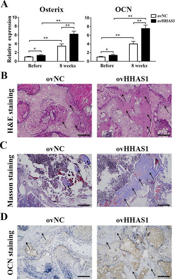
HHAS1 promotes BMSC osteogenic differentiation in vivo. BMSCs were loaded onto scaffolds and transplanted into nude mice. (A) The qPCR results showed that after implantation, the expression of Osterix and OCN increased obviously, and the HHAS1‐overexpression group displayed higher levels of these genes than the control group. (B) H&E staining showed more new bone formation in the HHAS1‐overexpression group (scale bar = 100 μm). (C) Masson's trichrome staining revealed that the HHAS1‐overexpression group had increased collagen organization (scale bar = 100 μm). (D) Immunohistochemistry showed that the HHAS1‐overexpression group displayed stronger staining of OCN (scale bar = 100 μm). The black arrows indicate the area of new bone formation. All experiments were performed three independent times, n = 5
