
Fig. 2.
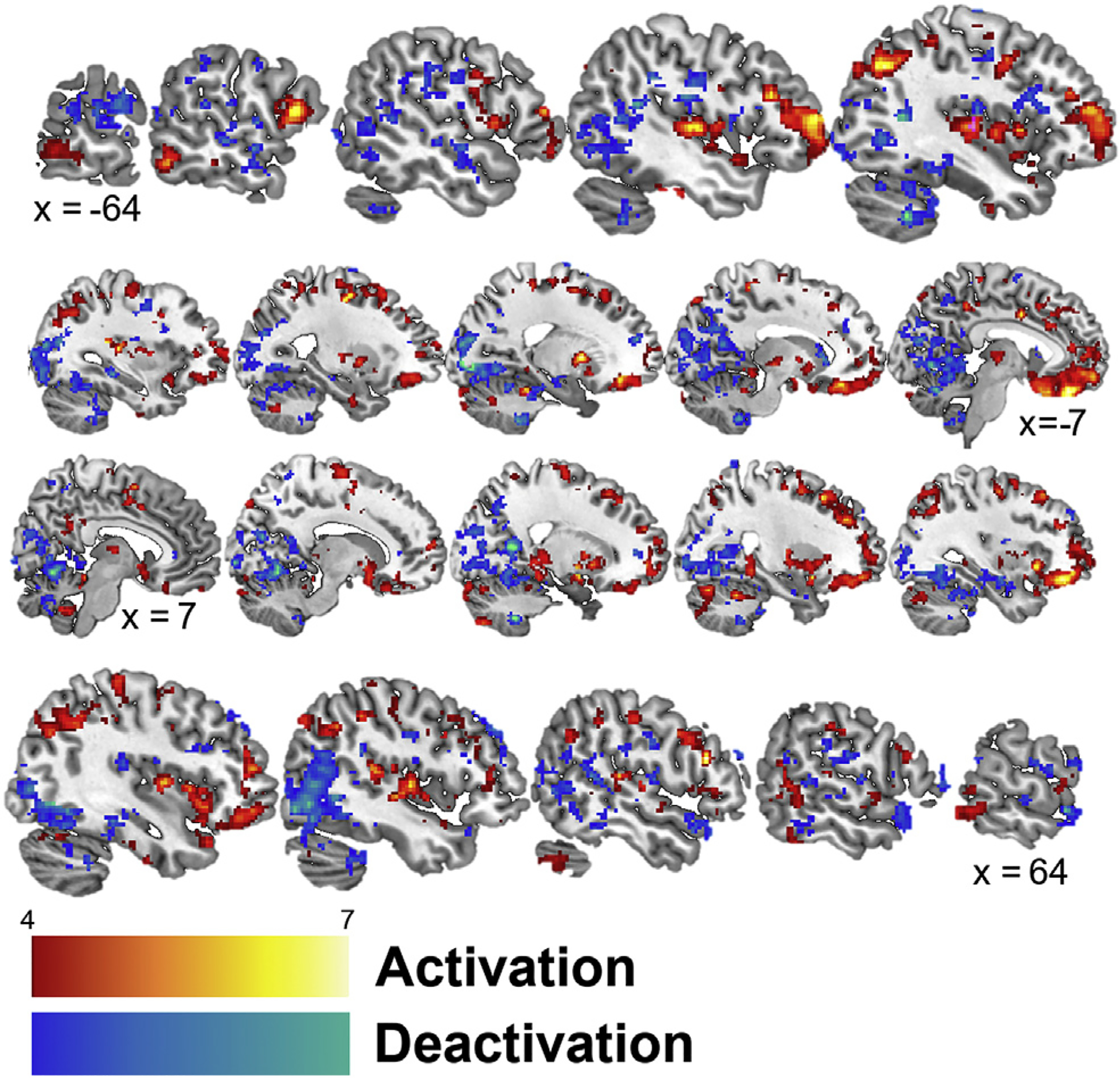
Sagittal slices presenting significant (p < 0.005) activation (red) and deactivation (blue) while listening to personalized trauma scripts in participants receiving cervical non-invasive vagal nerve and sham stimulation. Talairach x coordinates below slices indicate location with negative and positive coordinates located in the left and right hemisphere, respectively. Color bars indicate z-values of cluster.