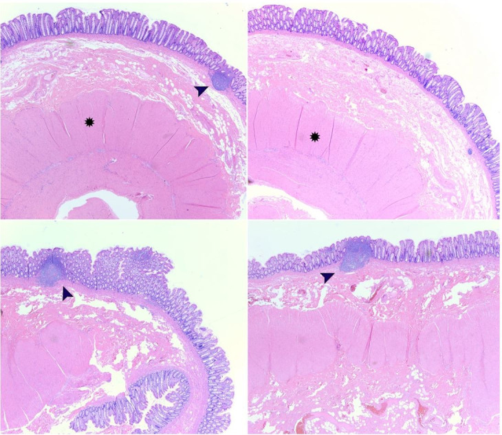
Figure 5. Microscopic examination using H&E stain. Histopathologic examination of the resected tubular colonic duplication demonstrates an intestinal wall containing all three layers (mucosa, submucosa, and serosa; true diverticulum) with well-formed smooth muscle layer (star) and mucosal lymphoid aggregates that extend to the submucosa (black arrows). Microscopic images were examined at 2.5x objective.

