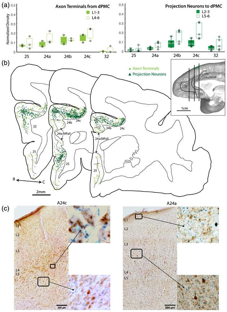
FIGURE 4.
Anterior cingulate cortex (ACC) connections with dorsal premotor cortex (dPMC). (a) Box and whisker and vertical scatter plots of normalized laminar density of tracer-labeled axon terminations from dPMC and projection neurons to dPMC. (b) Representative coronal maps (case PIM-FE tracer) of labeled projection neurons and terminations in ACC. Whole brain inset shows location of each slice along the rostro-caudal axis. (c) Representative coronal section photomicrographs of labeled dPMC projection neurons and terminations in A24c and A24a with Thionin counterstain. Low magnification images show pia to white matter, with laminar labels placed at the top of each layer. Insets are shown in higher magnification

