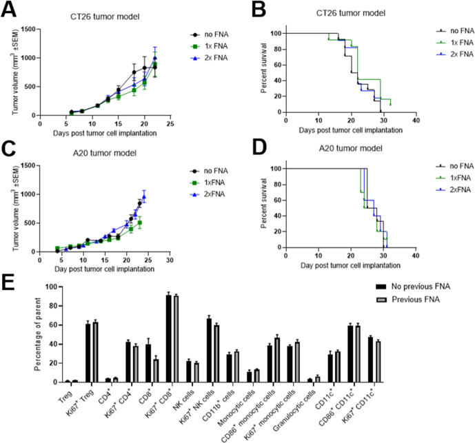
Figure 4.
Fine needle aspiration (FNA) biopsies can be carried out multiple times without impacting tumor growth or the tumor immune infiltrate. (A and B) Mice were subcutaneously implanted with CT26 tumors on the flank and the tumors were biopsied using FNA 12 days later (1× FNA group) or 12 and 14 days later (2× FNA group). Tumor growth over time (A) was compared between groups using a mixed effects model and time to endpoint (B) was compared between groups using a log-rank test. No significant differences were seen between FNA-biopsied and control groups. Error bars indicate the mean±SEM. Fourteen mice per group. The results represent one of three independent experiments. (C and D) Mice were subcutaneously implanted with A20 tumors on the flank and the tumors were sampled using FNA 15 days later (1× FNA group) or 13 and 15 days later (2× FNA group). Tumor growth over time (C) was compared between groups using a mixed effects model and time to endpoint (D) was compared between groups using a log-rank test. No significant differences were seen between FNA-biopsied and control groups. Error bars indicate the mean±SEM. Six to 10 mice per group. The results include data from three independent experiments. (E) Two to 3 days after FNA biopsying of CT26 tumors, the frequencies of 16 subsets of tumor-infiltrating immune cells were compared with those in mice that had not undergone FNA biopsying. For each subset, the groups were compared using a t-test with Holm-Sidak multiple comparison testing. No significant differences were seen between FNA-biopsied and control groups. Error bars indicate the mean±SEM. Five to 15 mice per group. The results include data from two independent experiments.

