
Figure 3. Polarized light images of hematoxylin and eosin–stained sections show growth factor dose-dependent changes in the tissue morphology (regions of interdigitation as noted by the arrows) at the repaired tendon-to-bone insertion site (Scale bar = 1 mm).
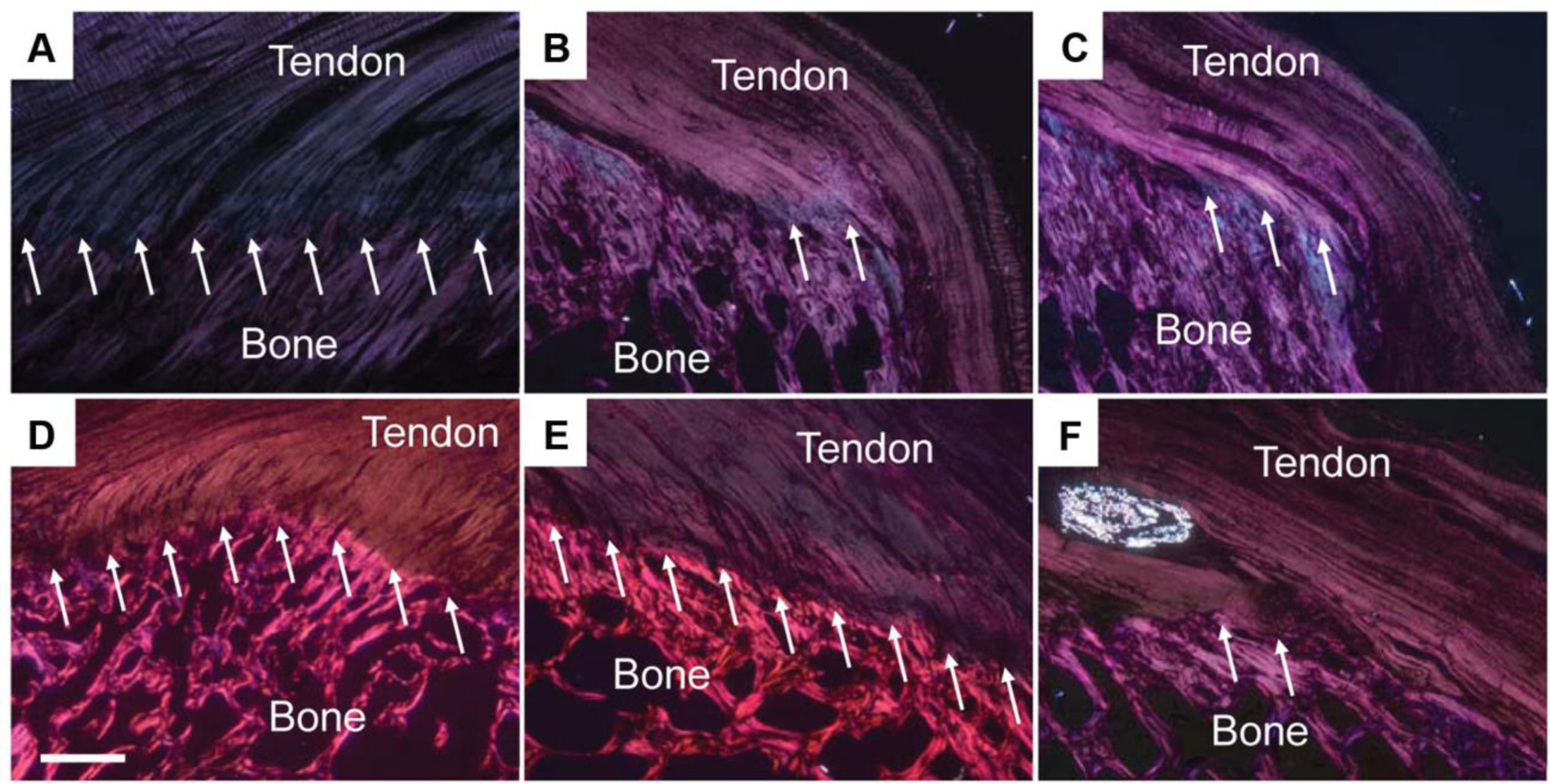
(A) Native tendon; (B) suture repair only; (C) collagen matrix+0 μg rhPDGF-BB; (D) collagen matrix+75 μg rhPDGF-BB; (E) collagen matrix+150 μg rhPDGF-BB; (F) collagen matrix+500 μg rhPDGF-BB. The morphological appearance was significantly improved in the 75 and 150 μg rhPDGF-BB augmented repairs when compared to the 0 and 500 μg rhPDGF-BB that showed poor morphology. Reprinted from Kee CH et al. Augmentation of a Rotator Cuff Suture Repair Using rhPDGF-BB and a Type I Bovine Collagen Matrix in an Ovine Model. The American Journal of Sports Medicine 2011; 39 (8):1630-39; with permission (SAGE publications).