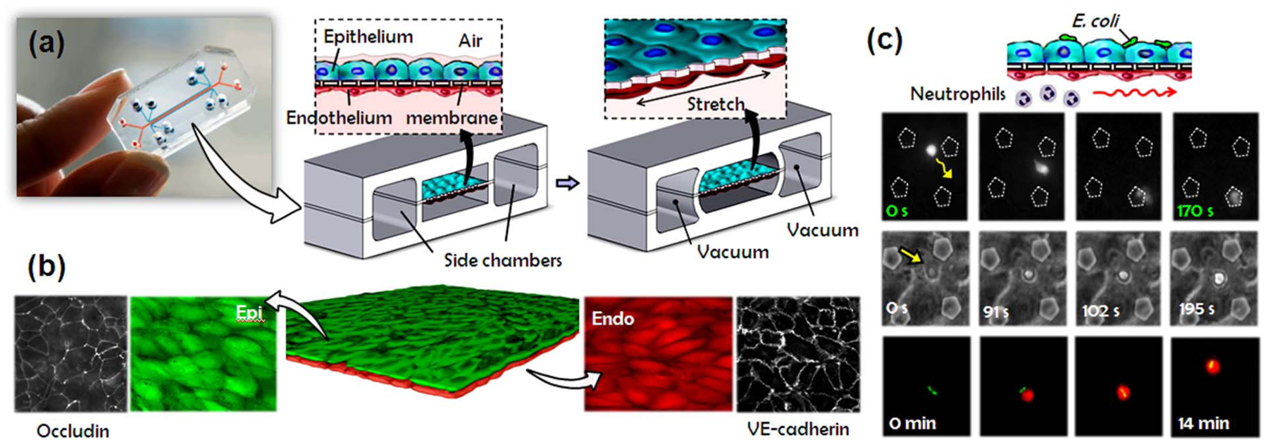
Fig. 5.

A human breathing lung-on-a-chip. (a) The microfabricated lung mimic device recreates physiological breathing movements by applying vacuum to the side chambers and causing mechanical stretching of the PDMS membrane forming the alveolar-capillary barrier. (b) Long-term microfluidic co-culture produces a tissue-tissue interface consisting of a single layer of the alveolar epithelium (Epi; green) closely apposed to a monolayer of the microvascular endothelium (Endo; red), both of which express intercellular junctional structures such as occludin or VE-cadherin. (c) Neutrophils flowing in the lower vascular channel adhere to the endothelium activated by E. coli in the alveolar chamber, transmigrate (top row), emigrate into the alveolar space (middle row), and engulf the bacteria (bottom row).
