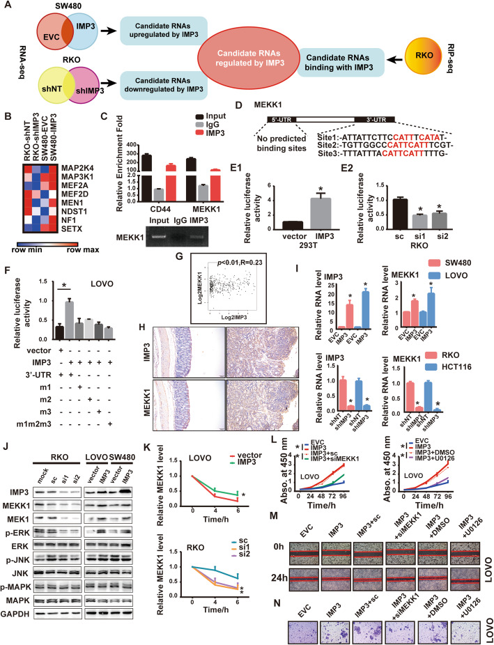
Fig. 4.
IMP3 directly binds to MEKK1 and activates MEK/ERK pathway. (A) Flow chart of bioinformatics analysis predicting candidate RNAs regulated by IMP3. (B) MAPK cascades were enriched in IMP3 regulated targets. (C) RIP assay showing that MEKK1 interacted with IMP3 in RKO cells. The RT-qPCR products were analyzed by electrophoresis (below) (*p < 0.05), CD44 was used as a positive control. (D) The schematic diagram of IMP3 potential binding site in MEKK1 UTR area. E-F. Luciferase reporter assay, (E1)293T cell was transfected with IMP3, (E2) RKO cell was transfected with IMP3 siRNAs, (F)LOVO cell was co-transfected with IMP3 and MEKK1 3’UTR wt and indicated mutations. The luciferase assay data were normalized to firefly luciferase activity. (*p < 0.05). (G)GEPIA analyzed the expression of IMP3 and MEKK1 in CRC tissues. (H) Representative images of IHC staining of IMP3 and MEKK1 in the colon tissues from Villin-Cre IMP3−/− and IMP3fl/fl mice treated with AOM/DSS. (I) The mRNA level of MEKK1 in indicated cells (* p < 0.05). (J) The protein level of MEKK1 and its downstream pathways in indicated cells. MEK1: mitogen-activated protein kinase kinase 1; ERK: extracellular-signal-regulated kinase; JNK: c-Jun N-terminal kinase; MAPK: mitogen-activated protein kinase; The prefix p represents its phosphorylation. (K) RT-qPCR analysis of MEKK1 mRNA stability in actinomycin D treated CRC cells (* p < 0.05). (L) CCK-8 results showed that knockdown of MEKK1 or using MEK/ERK inhibitor U0126 attenuated the enhanced cell viability induced by overexpression of IMP3 in LOVO cells (* p < 0.05). Representative images of (M) wound healing assays and (N) transwell assays showed that MEKK1 repression or U0126 rescued the enhanced invasion and migration ability by overexpression of IMP3 (* p < 0.05)

