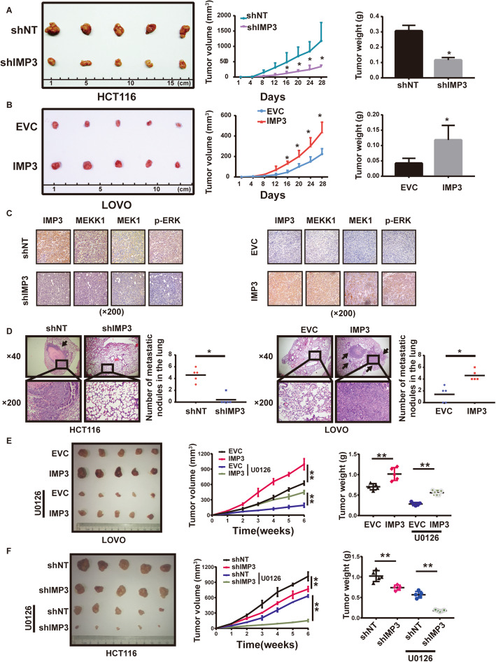
Fig. 5.
IMP3 promotes CRC cell proliferation and metastasis in vivo. (A-B) 2 × 10⁶ stable cells were injected subcutaneously into the groin of nude mice (n = 5 for either group). (A) Knockdown of IMP3 declined tumor growth and tumor weights compared with those control cells in HCT116 (B) Overexpression of IMP3 promoted tumor growth and tumor weights compared with those control cells in LOVO. (C) Representative images of IHC staining for IMP3, MEKK1, MEK1 and p-ERK. (D) Representative images of lung metastasis in nude mice with HE staining. (left) Knockdown of IMP3 reduced the number of lung metastasis nodular compared with those control cells in HCT116, (right) Overexpression of IMP3increased the number of lung metastasis nodular compared with those control cells in LOVO (* p < 0.05). (E) Representative image of nude mice bearing tumors formed by overexpression of IMP3 in LOVO and their control cells after U0126 treatment. The average tumor volume and tumor weight of nude mice bearing tumors formed by overexpression of IMP3 in LOVO and their control cells after U0126 treatment. (* p < 0.05, ** p < 0.05) (F) Representative image of nude mice bearing tumors formed by stable knockdown of IMP3 in HCT116 and their control cells after U0126 treatment. The average tumor volume and tumor weight of nude mice bearing tumors formed by stable knockdown of IMP3 in HCT116 and their control cells after U0126 treatment. (* p < 0.05, ** p < 0.05)

