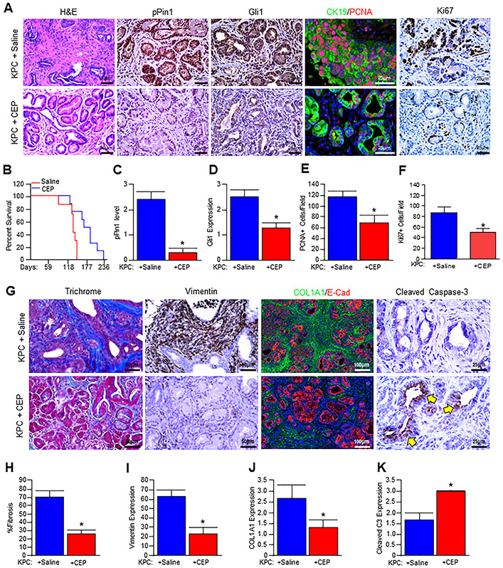
Figure 6. CEP-1347 reduces pancreatic tumor burden and extends survival in vivo.
(A) Pdx1-Cre x LSL-KRASG12D x LSL-TP53R172H (KPC) mice at 3 months of age were treated daily either with saline (vehicle) or 1.5 mg/kg CEP-1347 for seven weeks. At the conclusion of the study, pancreatic tissues from control and treated mice were stained with H&E or via immunohistochemistry or immunofluorescence for PIN1-pS138, GLI1, or CK19/PCNA, or Ki67. (B) Kaplan-Meier curve indicating survival for control (saline) and CEP-1347 treated mice. (C-F) Tissue sections were scored by two investigators and quantified for the indicated proteins. Average values are presented as mean ± SEM, and two groups were compared by the two-tailed Student t-test (N=3, *p<0.05). (G) Pancreatic tissues were next stained with either Masson’s trichrome or via immunohistochemistry or immunofluorescence for Vimentin, COL1A1/E-Cad, or apoptosis surrogate, Cleaved Caspase-3. (H-K) The stained sections were quantified, and average values are presented as mean ± SEM, and two groups were compared by two-tailed Student t-test (N=3, *p<0.05) and presented as mean ± SEM.

