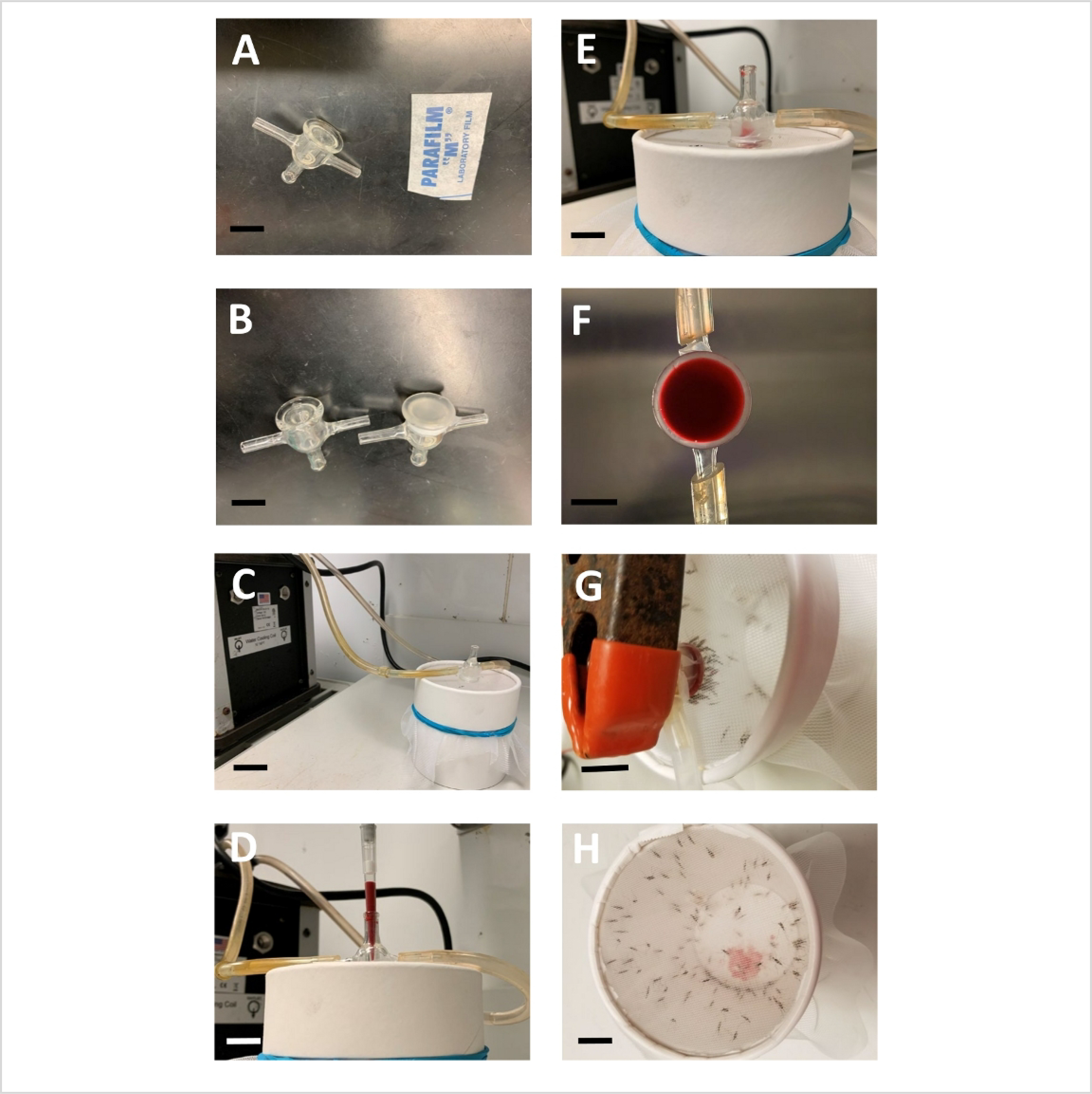
Figure 2: Mosquito blood feeding set-up.

(A) Glass feeder and rectangular piece of paraffin film (B) Two glass feeders displayed before and after parafilm membrane attachment (C) Glass feeder on top of mosquito cup and connected with circulation water bath. (D,E) Pipetting of blood feed into the glass feeder (F) Bottom view of glass feeder showing homogenous distribution of blood feed (G) Several mosquitoes feeding through parafilm membrane. (H) Top view of mosquito cups after feeding, showing drops of blood excreted by the feeding mosquitoes at the bottom of cup. Scale bar = 10 mm.
