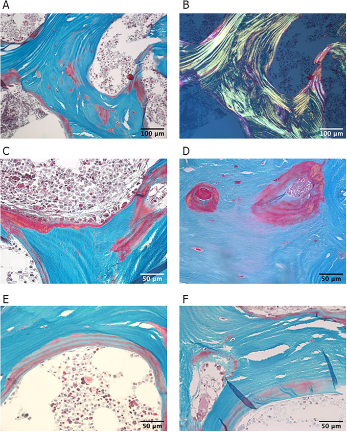
Fig. 1.

(A) Bright‐field light microscopy images of a thin section (3 μm) of trabecular bone stained with Goldner trichrome (green corresponds to mineralized bone matrix, orange/red to non/mineralized bone matrix—osteoid). (B) Corresponding polarized light microscopy shows regular lamellar organization. Note the presence of nonmineralized bone tissue in red in the central part of the trabeculae. (C–F) Bright‐field light microscopy images of higher resolution showing regions with a large amount of osteoid (C, D). (E, F) Osteoid with interlacing regions stained in green and red, suggesting part mineralization.
