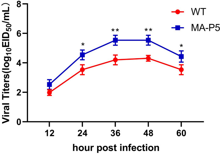
Figure 2.
To characterize the growth kinetics, MDCK cells were infected with WT or MA-P5 at an MOI of 0.01 TCID50 per cell and treated with 1 µg/mL TPCK. The MDCK cell culture supernatants were harvested at 12, 24, 36, 48 and 60 dpi and stored in a -80°C freezer. The titers were calculated to determine the TCID50 at every time point by the Reed–Muench method. *P < 0.05 (MA-P5 vs WT), **P < 0.01 (MA-P5 vs WT). To determine the virulence of WT and MA-P5, mice (n = 5) were intranasally inoculated with 106 EID50 of WT and MA-P5. An equal volume of PBS was used as a negative control.

