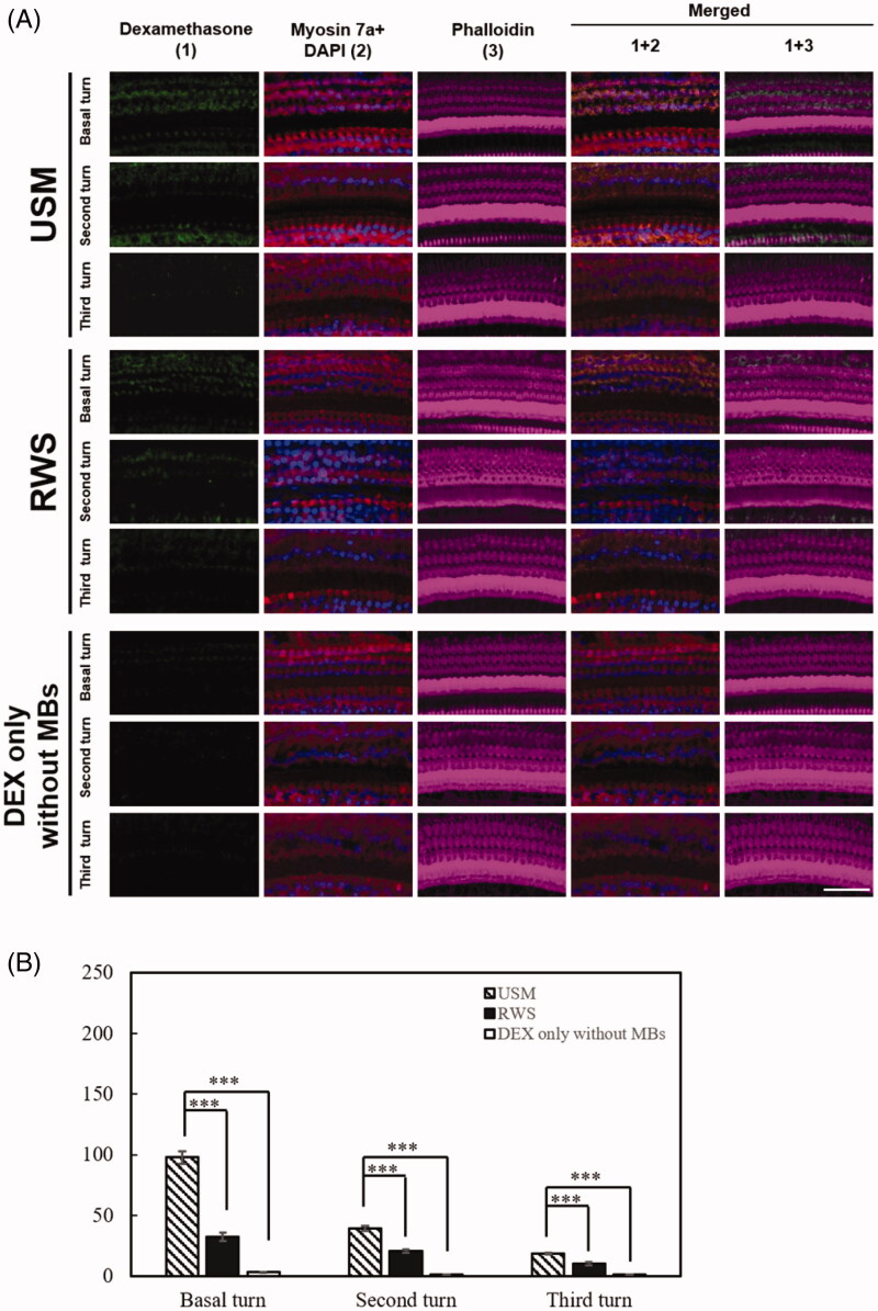
Figure 9.
Exposure to the US-mediated P407-MB gel maintains the cellular uptake of DEX in the cochlea for one week. Samples were obtained from the cochleae 7 days after DEX treatments. (A) Representative confocal microscopic images of indirect immunofluorescence staining show stronger localization of DEX uptake (green) in the USM group than in the groups with RWS and DEX only without MBs in the cochlea. Four repetitions of this experiments were conducted. Myosin 7a, cell bodies (red); phalloidin, stereocilia bundles (magenta); DAPI, nuclei (blue). (B) Histogram representations of the mean fluorescence intensity of DEX staining. Data are shown as the means ± SEM (n = 4 for each bar). Scale bar: 50 μm. USM, ultrasound microbubble treatment; RWS, round window soaking; MBs, microbubbles; DEX, dexamethasone.

