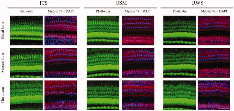
Figure 11.
Representative images of confocal microscopic immunofluorescence analysis of cochlear surface preparation 28 days after USMB treatments. There was no difference in the survival of cochlear hair cells between the USM, RWS and ITS groups. The staining shows the nuclei (blue, DAPI), filamentous actin (green, phalloidin), and cell bodies (red, myosin 7a). Four repetitions of this experiments were conducted. Scale bar: 50 μm.

