
Figure 6.
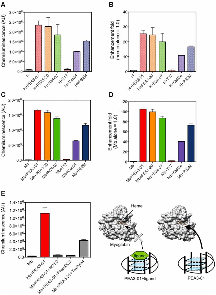
PEA series upregulated peroxidase activity of hemin, and hemin-binding aptamers enhanced chemiluminescent signal from luminol reaction catalyzed by myoglobin. T17 was used as a negative control. All measurements were performed in triplicate except for the data of hemin-T17 and hemin-CatG4 (one experimental result was lost for each) and the data are shown as mean ± SD in the graph. (A–B) 100 nM Hemin (H) was incubated with 200 nM PEA3-01, PEA1-20, N24-07, T17, CatG4 or PS2M in potassium acetate buffer (pH 5.0). Enhancement of the chemiluminescent signal corresponding to luminol substrate oxidation was evaluated (A). Relative enhancement fold (hemin alone = 1.0) was shown in (B). (C–D) Enhanced chemiluminescent signals with hemin-binding G4 aptamers, CatG4 and PS2.M. 100 nM myoglobin (Mb) was incubated with 200 nM PEA3-01, PEA1-20, N24-07, T17, CatG4 or PS2M in potassium acetate buffer (pH 5.0). Chemiluminescent signal (C) and relative enhancement (myoglobin alone = 1.0) (D) are shown. (E) Competitive assay using G4 ligands. Activity measurement was performed using 100 nM PEA3-01, 0 or 500 nM G4 specific ligands, and 100 nM myoglobin.